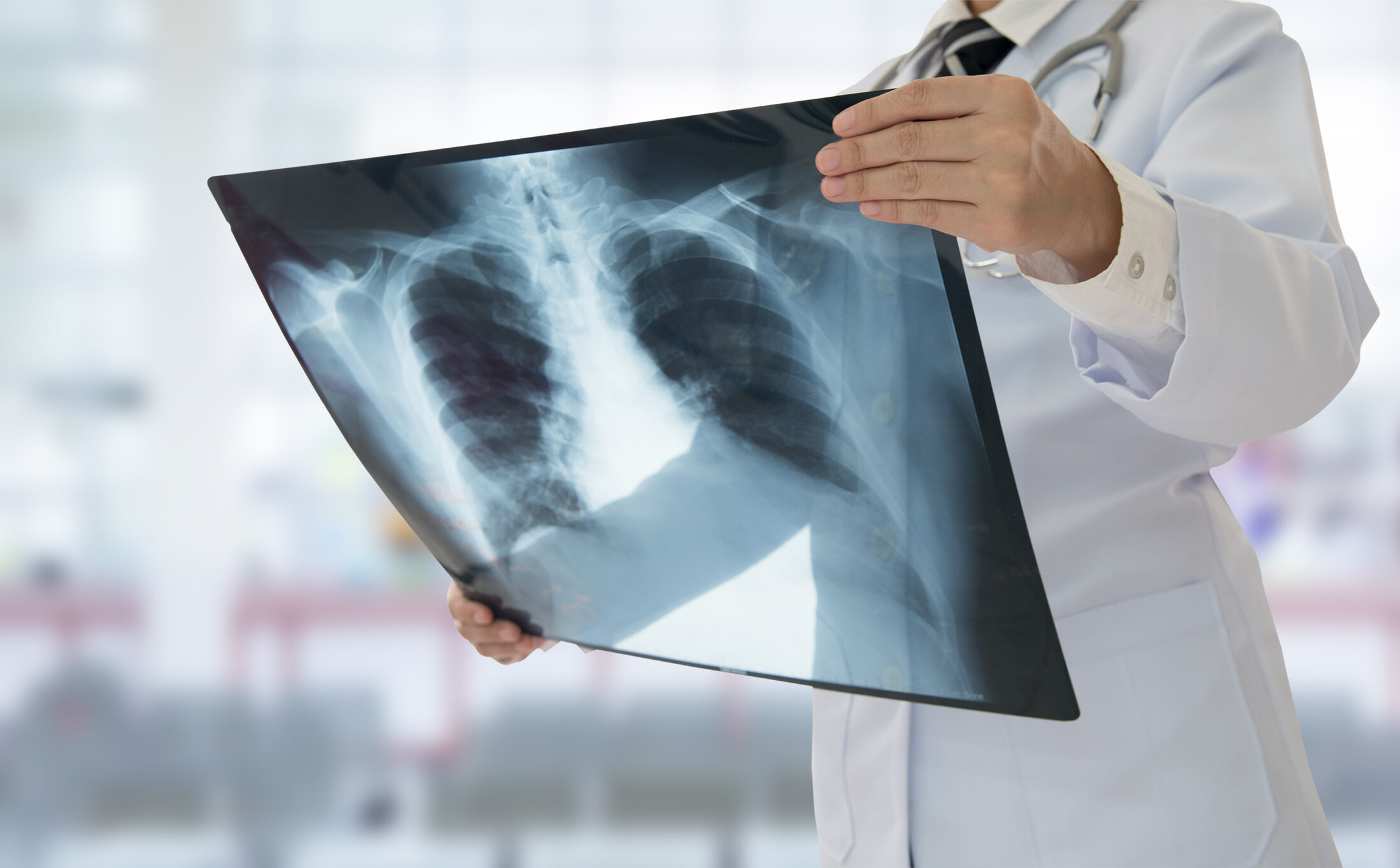
Radiograph (X-ray)
Radiography (x-ray) is a diagnostic imaging test. Low dose radiation is used with a detector to generate pictures of the body. Radiologists will then review the images to help diagnose various conditions. This test is useful for diagnosing bone or joint abnormalities, to evaluate the lungs, and to evaluate for kidney stones among other reasons. The process of undergoing an x-ray is quick and requires minimal preparation. Radiation dosage is low and research shows that the value of x-ray imaging outweighs the risks of radiation exposure.
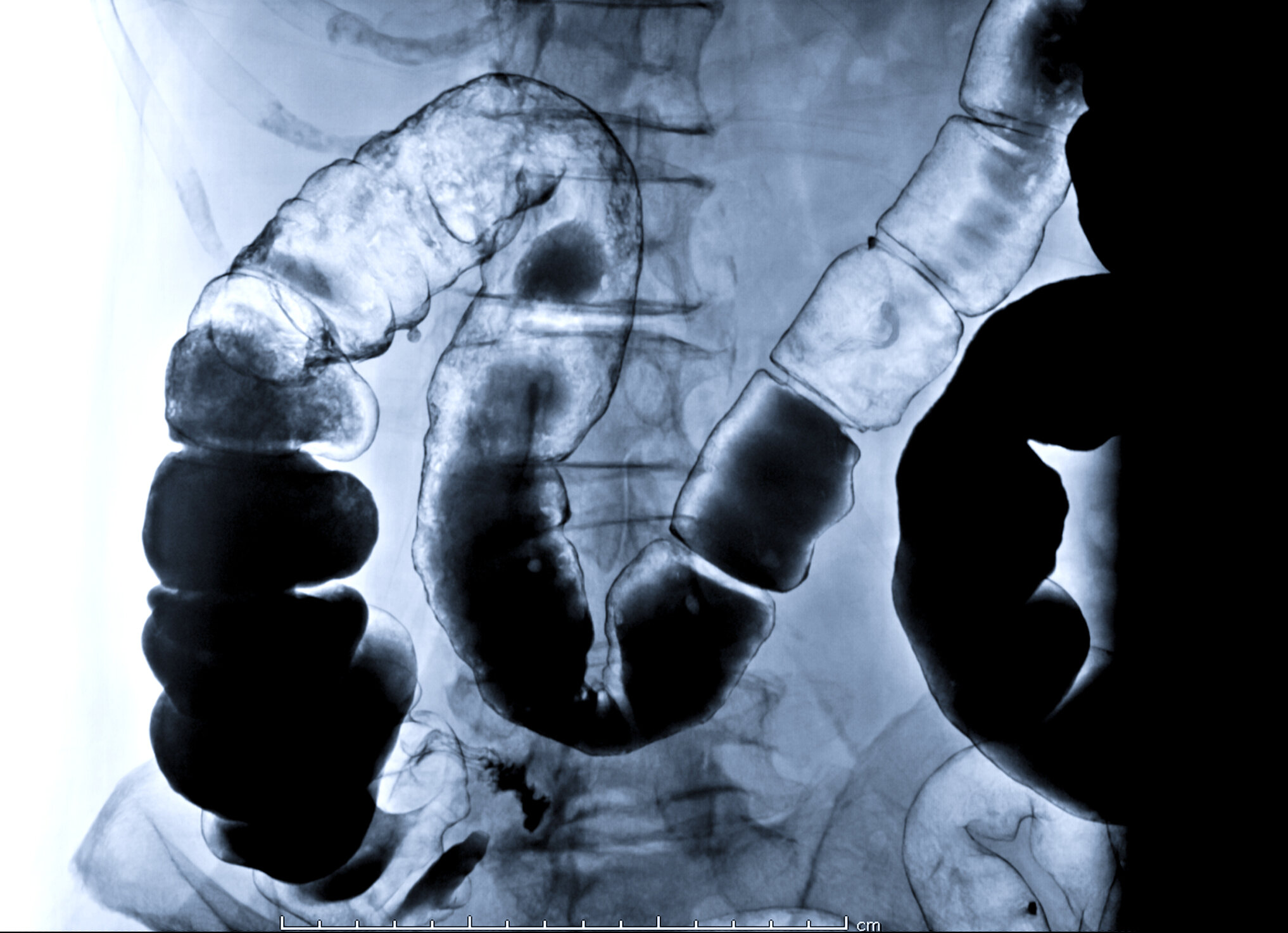
Fluoroscopy
Fluoroscopy describes an imaging technique in which real time x-ray video is used to diagnose and in some cases treat medical conditions. Such studies include imaging the intestinal tract, the urinary system, and to provide image guidance during pain intervention procedures. There is variable length to a fluoroscopic exam depending on the patient. Specific instructions including what to expect and any special preparation required such as fasting will be communicated to you prior to your fluoroscopy exam.
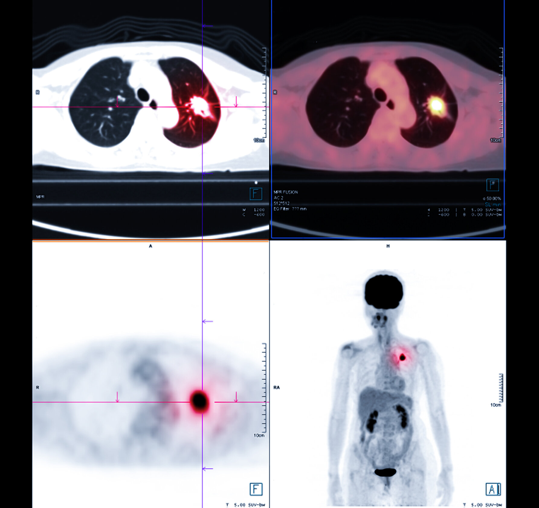
Nuclear Medicine
Nuclear medicine describes a broad range of imaging studies and therapies that involve administering radioactive medicines. These materials travel to the organ system of interest and emit energy which can treat some medical conditions or be detected by a special camera to produce an image. Nuclear medicine is often used to assess heart function or to evaluate cancer (PET Scan). In some cases, nuclear medicine is used treat thyroid problems and cancer.
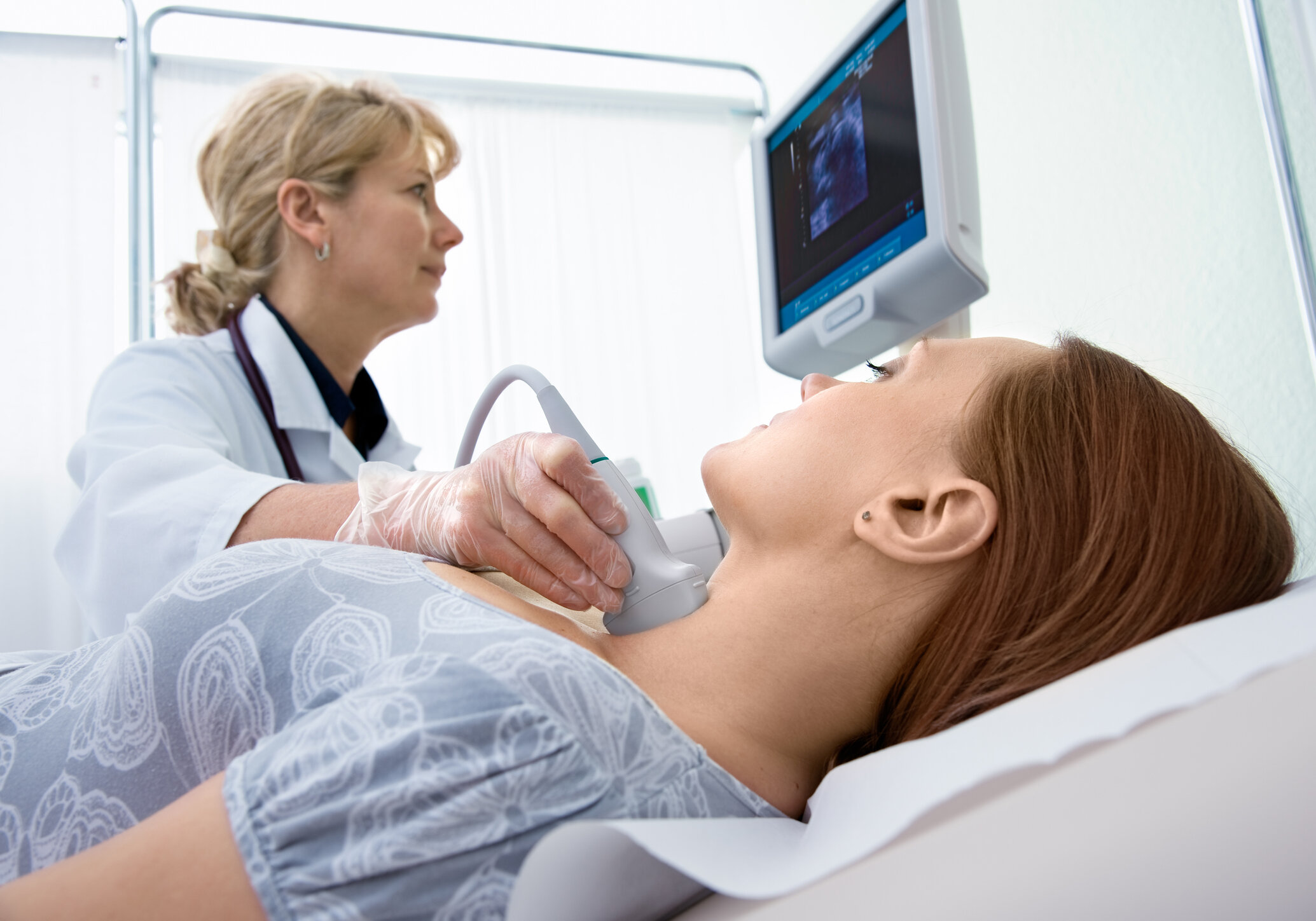
Ultrasound
Ultrasound, also known as sonography, uses a transducer placed on the body to emit sound waves and generate images. It is useful to evaluate soft tissues or hollow structures like the gallbladder or blood vessels. Ultrasound is also utilized to guide radiologists as they perform certain procedures such as a biopsy, abscess drainage, or joint injection. Ultrasound does not use radiation so it is safe and useful to evaluate pregnancies. In some cases special preparation such as fasting may be required prior to the examination.
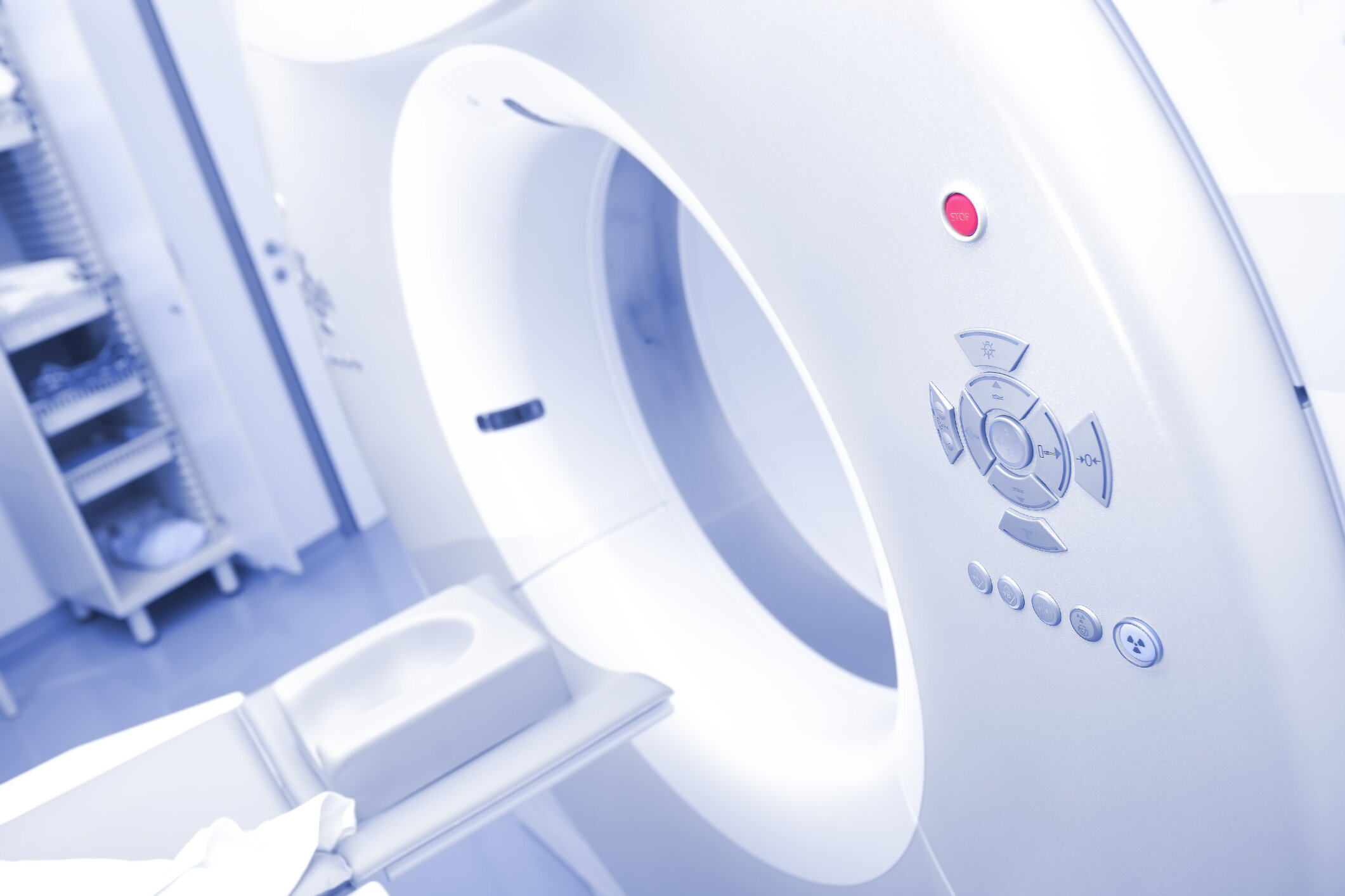
Computed Tomography (CT)
Computed tomography (CT scan) is a diagnostic imaging test that uses radiation and a computer to generate cross sectional pictures of the body and internal anatomy in high detail. CT can show bone and soft tissue structures such as internal organs, muscles, and blood vessels. It can be utilized to diagnose a wide variety of medical conditions. A CT scan may require a contrast media to better highlight abnormalities and assist the radiologist with making the most accurate diagnosis. These studies are performed by certified imaging technologists, and every examination is directly supervised and interpreted by our board certified radiologists.
Magnetic Resonance Imaging
What is an MRI?
Magnetic resonance imaging (MRI) is one of the most accurate ways to examine soft tissues, organs, and bones. It is a safe and painless examination that does not use radiation. Instead, MRI combines computer technology with magnets to create very clear, highly defined images of the area of interest. MRI is often considered the “gold standard” for imaging specific organs such as the liver or kidneys.
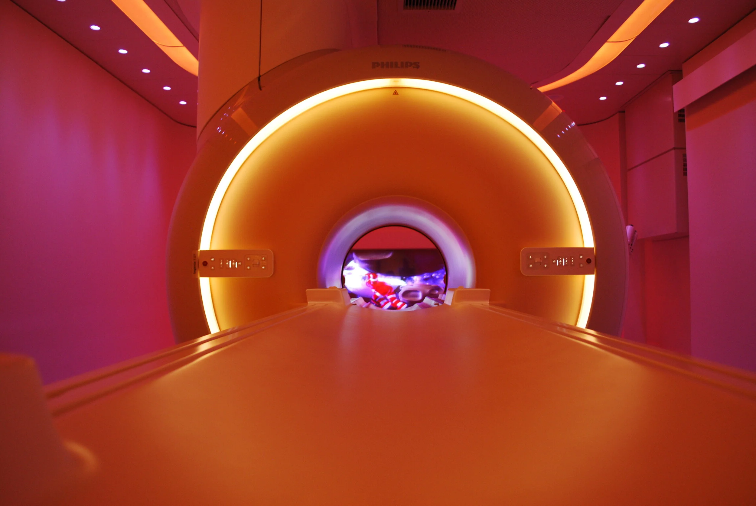
The Experience
Lahey Outpatient Center in Danvers is the first in the U.S. to offer the state-of-the-art Ambient MRI Experience – the most soothing patient environment available for MRIs while giving physicians high-quality imaging results.
Ambient MRI has been clinically proven to decrease anxiety and increase patient well-being. Patients can choose their own music, lighting and wall images to transform the wide and spacious MRI suite into a soothing multi-dimensional theater.
The MRI scanner itself offers the widest oval open design on the market. The Ambient Experience is especially recommended for patients with claustrophobia, anxiety and painful medical conditions who will benefit from a spacious and more comfortable MRI examination.
We invite you to watch a short video that highlights this innovative MRI experience:
Designed for Patient Comfort
Wide and open design to ease stress and anxiety
Interactive lighting, projection and sound for a comfortable and relaxing atmosphere
Various projected themes, soothing music and dynamic, colored lighting
Advanced noise reduction technology that minimizes vibrations and noise
Faster scanning time
Feet-first scanning with your head outside of the scanner for 70% of exam types
Accommodates patients up to 550 lbs.